


Varicose veins during pregnancyare an area of venous vessels that appeared during pregnancy and are pathogenetically related to it. It is manifested by severity, paraesthesia, pain in the lower extremities and external genitalia, swelling, muscle contractions, nutritional lesions of the skin. It is diagnosed by examination, methods of ultrasound angiography. During pregnancy, treatment is usually limited to compression therapy with sleep and rest correction, physical activity and diet. Perhaps the appointment of phlebotonics, venoprotectants, anticoagulants, antiplatelet agents. Surgical treatments are usually used after childbirth.
General information
Varicose veins (varicose veins) are one of the most common vascular diseases associated with pregnancy. According to studies, up to 15-20% of people suffer from venous pathology, while 2/3 of them are women and 60-80% of venous cases have occurred due to pregnancy. The disease is usually first diagnosed in young patients, 75% of whom are under 30 years old. In more than two-thirds of cases, the varicose vein clinic begins after the 20th week of the first pregnancy. The relevance of early diagnosis of varicose veins is related to the high probability of fetal insufficiency and the risk of fatal thromboembolic complications in the absence of appropriate treatment.Reasons
Given the statistics on the incidence of varicose veins during pregnancy, most obstetricians and gynecologists consider the disease as a complication of pregnancy. The predisposing factor that causes vascular dilatation in 91% of patients is a genetically determined median venous insufficiency in which the amount of collagen decreases and the polysaccharide content increases. The development of varicose veins in constitutionally predisposed women during pregnancy is facilitated by:
- Increased volume of circulating blood. The increase in BCC in pregnant women ranges from 30-50% (when carrying a child) to 45-70% (if there are 2 or more fetuses in the womb). This compensatory mechanism ensures an adequate blood supply to the child, the woman's vital organs and the fetal placental system.
- Hormonal adjustment during pregnancy. During pregnancy, the ovaries and placenta secretively secrete progesterone and relaxation. Under the influence of these hormones, the smooth muscle fibers of the veins relax and the structural reconstruction of the connective tissue takes place. As a result, the vascular wall responds worse to increased intravenous pressure.
- Vascular compression of the pregnant uterus. The developing uterus compresses the inferior vena cava and the iliac vein. Blood flow from the pelvis and lower extremities is reduced, intravascular pressure is increased, which causes the venous walls to stretch. The effect of this factor plays a key role in the formation of varicose veins after the 25th week of pregnancy.
- Changes in the hemostasis system. As labor approaches, the fibrinolytic activity of the blood decreases and the number of coagulation factors increases. This adjustment mechanism aims to reduce the volume of normal blood loss during labor. This increases the chance of thrombosis of abnormally damaged veins.
Pathogenesis
The starting point in the development of varicose veins during pregnancy is the cessation of the compensatory capabilities of the venous valve device. Due to the increase in BCC and mechanical obstruction in the outflow from the lower extremities, when the main veins are compressed, the blood exerts increased pressure on the vascular wall. Genetic hereditary failure of connective tissue fibers is enhanced by the relaxation of vascular smooth muscle under the action of progesterone. As a result, the lumen of the vein dilates, the valves stop closing, blood is deposited in the vascular system of the lower extremities. As the disease progresses, the pathological process can spread to the vessels of the vulva, vagina and small pelvis.
Classification
The main criteria for the systematization of varicose veins are the anatomical prevalence of venous stasis and the severity of the disease. This approach allows a differentiated choice of treatments for different variants of the disorder. Taking into account the involvement of various organs in the process, the varicose veins of the lower extremities, the varicose veins, the varicose veins of the pelvic organs can be distinguished. Depending on the severity of the clinical symptoms, the following stages of venous dilatation of the lower extremities can be distinguished:
- Compensated varicose veins. There are no external signs of vasodilation, the pregnant woman notes fatigue of the legs by the end of the day, discomfort in the calf muscles during exercise and brisk walking.
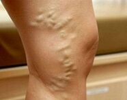
- Damaged varicose veins. A vascular pattern ("stars") appears on the skin. At night, the legs swell, at night there are cramps, numbness, pain. Bruises and scratches heal more than usual.
- Compensated varicose veins. The patient is constantly worried about leg pain, swelling increases. The veins have enlarged, knots. The skin is hyperpigmented. There are signs of eczema and eating disorders.
With pelvic varicose veins in pregnant women, the disease also develops gradually. In the first stage, the diameter of the affected vessels in any venous plexus of the pelvis does not exceed 5. 0 mm. With the second, the uterus or ovaries are involved in the process, the lumen of the vessels is 6, 0-10, 0 mm. The third is characterized by venous ecstasy over 10 mm with full involvement of all pelvic venous plexuses.
Symptoms of varicose veins
In 80-82% of patients, the disease begins with a feeling of heaviness, tension, "buzzing" in the legs, increases at night and during exercise. The symptoms of varicose veins are gradually increasing. As the disease progresses in certain areas of the muscles, pain appears, which initially develops with prolonged posture, performing physical work. In the most severe cases, the pain becomes constant and its intensity can be so intense that the pregnant woman has difficulty moving independently. Up to 60% of patients experience cramps in the muscles of the calf, up to 40-50% - loss of sensation, numbness of the legs, up to 30% - itching.
In the varicose vein compensation stage, external signs of superficial vein dilatation appear. First, areas of reticular vessels and telangiectasias ("lattice" and "stars") form on the skin. The venous pattern then becomes distinct. The veins appear dilated, complex and nodular over time. The spread of the extension to the deep vessels is evidenced by the appearance of swelling in the ankle joints and lower legs. With the decompression of the varicose veins, the skin of the feet looks hyperpigmented, eczema develops. If the pathology arose long before pregnancy, subcutaneous adipose tissue dystrophy, food ulcers are possible.
In 4% of patients, the disease affects the veins of the vulva, vagina and small pelvis. With venous and vaginal varicose veins, discomfort, distension, gravity, itching are observed in the area of the external genitalia. There may be swelling of the perineum and lips, contact with vaginal bleeding after sex. Pelvic congestion syndrome is manifested by pulling or aching pains in the lower abdomen, which radiate to the lower back, sacrum, groin and external genitalia. Dyspareunia (pain during intercourse) is typical. In severe cases, dysuric disorders are detected.
Complications
In the absence of appropriate treatment, varicose veins in pregnant women can be complicated by the development of trophic ulcers, erysipelas, thrombophlebitis, superficial and deep vein thrombosis, pulmonary artery thromboembolism and other pulmonary arteries. In 40-45% of cases, placental insufficiency occurs with acute and chronic fetal hypoxia. In 25% of patients, abnormalities at work are observed (weakness of labor force, discrepancy of the contractile activity of the myometrium). With atrial fibrillation, massive traumatic course of the postpartum period is possible. Nearly one-third of working women have defects in placental abruption and placental abruption. The long-term consequences of varicose veins that occur during pregnancy are hemorrhoids, chronic venous insufficiency and pelvic pain.Diagnostics
With the appearance of characteristic spots on the skin, the diagnosis of varicose veins during pregnancy usually presents no difficulties. The tasks of the diagnostic stage are to determine the stage and the location of the venous extension, to exclude other causes that can cause stagnation in the vascular system of the lower extremities. The most informative research methods are:
- Chair inspection. The study reveals characteristic changes in the venous vessels in the area of the vulva and in the inner thighs - stretch, turtle, nodular. Swelling of the lips and perineum is possible. When viewed in mirrors, the vaginal mucosa appears hypertrophic, cyanotic. Atrial fibrillation is normalized by bilateral palpation, often painfully.
- USDG of the venous system. When scanning with ultrasound, the shape and diameter of the vessels, their length, anatomical position and the condition of the wall are assessed. The method allows you to determine the junction zones, the coherence of the valve device, the sharpness of the veins, the presence and direction of the regression. You can scan both the vessels of the lower extremities and the inferior vena cava (IVC ultrasound).
- Double scan of leg vessels. The advantage of the non-invasive method, which combines traditional ultrasound and Doppler studies, is not only the obtaining of detailed information on blood flow parameters, but also the visualization of the venous network. Duplex angioscanning is used for a comprehensive assessment of the condition of superficial, perforated and deep vessels.
Radiodiagnostic methods (varicose veins, selective ovulation, ascending venipuncture, pelvic venography, CT venography, venoscopy, etc. ) during pregnancy are used to a limited extent due to possible adverse events. In difficult cases, with suspected pelvic varicose veins, diagnostic laparoscopy is performed with caution. The differential diagnosis of varicose veins of the legs is made with a drop of pregnant women, heart failure, lymphedema, acute venous thrombosis. Varicose veins of the small pelvis should be differentiated from endometriosis of the genitals, chronic inflammatory pathology of the pelvic organs, subcutaneous and underwater uterine myomas, cysts and other ovarian tumors. In addition to observing an obstetrician-gynecologist, the patient is advised to consult a venologist, cardiologist and oncologist.
Treatment of varicose veins during pregnancy
The main goals of treatment for varicose veins in pregnant women are to stop the development of the disorder, to alleviate the severity of the clinical picture and to prevent possible thromboembolic complications. Non-pharmacological methods are considered preferable, if necessary, supplemented with pharmacotherapy in safe periods of pregnancy:
- Compression therapy. It is recommended for a woman with a confirmed diagnosis of varicose veins every day throughout pregnancy, to use elastic bandages, especially compression tights or tights of 1-2 degrees of compression during childbirth and the postpartum period. Compression therapy by mechanically reducing the diameter of the superficial veins accelerates blood flow, reduces swelling and congestion.
- Plant-lighting and veniprotective plants. The effect of the use of drugs of this group is associated with an increase in the tone of the venous wall, reduction of its permeability, improvement of microcirculation, rheological properties of blood and lymph outflow. The advantage of most bioflavonoids is that they can be used during pregnancy and lactation. Phlebotonic drugs are prescribed both in tablet form and externally.
- Anticoagulants and antiplatelet agents. In the presence of signs indicating a tendency for increased coagulation and the threat of DIC development, anticoagulant drugs are used with caution. To improve the rheology of the blood and the vascular microcirculation, there are drugs that prevent the accumulation of platelets and have a vasoprotective effect.
Recommended to pregnant women with varicose veins special complexes of physiotherapy exercises, lymphatic drainage massage, dosimetric walking, daily ascending contrast shower. Diet correction includes eating foods rich in fiber and vegetable fats. Injection sclerotherapy, miniblectomy, cross-section, intranasal laser coagulation and other surgical treatments are used in exceptional cases with severe forms of the disease, severe pain syndrome and the presence of complications. Most often, surgical correction is performed at the end of the lactation period.
Delivery tactics
The preferred delivery method for varicose veins is natural childbirth, at the beginning of which elastic bandages or compression garments are applied to the lower extremities of the woman. Patients with vulvar-vaginal varicose veins require particularly careful maintenance of the persistent period by performing protective perineotomy, as indicated. When the veins rupture, the damaged vessels are carefully ligated with repetitive sutures of the group. Cesarean section is recommended in patients at high risk of thromboembolic complications and severe varicose veins.
Prediction and prevention
With early detection and adequate treatment, the prognosis is favorable. For prophylactic purposes, it is recommended that you get adequate night's sleep and periodic rest throughout the day in a supine position with your feet placed on a stable surface at a 30 ° angle. Pregnant women with severe heredity should refuse to wear shoes with heels over 5 cm, limit the duration of sitting or standing and control weight gain.
To prevent varicose veins, daily walking, reducing salt intake, taking vitamin supplements that strengthen the vascular wall are effective. Patients with varicose veins who are planning a pregnancy, if appropriate, undergo surgery to correct the disease.